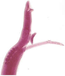
Schistosoma mansoni


We have recently focused on the use of the mouse peritoneal cavity for studying the cercaria - schistosomule transformation as well as the effect of drugs on such transformation. During these studies, it was observed under stereomicroscope, that the living recovered larvae exhibited host cells adhered to their surfaces. So the kinetic of cell adhesion to Schistosoma mansoni larvae developing in the peritoneal cavity of naive mice was investigated, and the phenomenon was further studied by light, transmission and scanning electron microscopies. On the other hand, cercariae of S. mansoni after inoculation into the peritoneal cavity of AKR/J strain of mice developed and reached the sexual maturation in that site with egg prodution. No evidence of hemoglobinic pigment in their digestive tract was observed in the peritoneal recovered parasites from both strains (AKR/J and SWISS). This fact suggested that the parasites can develop withouth red blood cells ingestion. On the other hand, the development of the parasite with egg prodution in the peritoneal cavity of the AKR/J mouse reinforces the data that the lung phase is not necessary for the development of the parasite.